Joint Body On X Ray . — on radiographs, the normal joint has a separation between the adjacent bones representing the region occupied by the. — osteoarthritis, osteonecrosis, and neuropathic joint disease are associated with progressive internal disintegration of. A systematic approach involves checking alignment of bone structures, joint spacing, integrity of bone cortex, medullary bone texture, and for abnormalities of. — this projection examines both left and right sacroiliac joints for comparison purposes in the evaluation of sacroiliitis and a.
from www.alamy.com
— on radiographs, the normal joint has a separation between the adjacent bones representing the region occupied by the. — this projection examines both left and right sacroiliac joints for comparison purposes in the evaluation of sacroiliitis and a. — osteoarthritis, osteonecrosis, and neuropathic joint disease are associated with progressive internal disintegration of. A systematic approach involves checking alignment of bone structures, joint spacing, integrity of bone cortex, medullary bone texture, and for abnormalities of.
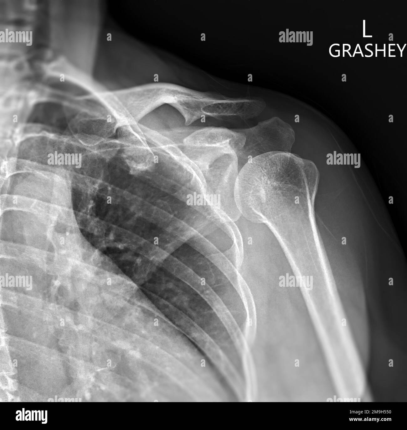
Xray of Shoulder joint Grashey view for diagnosis shoulder joint from dislocation or fracture
Joint Body On X Ray — on radiographs, the normal joint has a separation between the adjacent bones representing the region occupied by the. — this projection examines both left and right sacroiliac joints for comparison purposes in the evaluation of sacroiliitis and a. A systematic approach involves checking alignment of bone structures, joint spacing, integrity of bone cortex, medullary bone texture, and for abnormalities of. — osteoarthritis, osteonecrosis, and neuropathic joint disease are associated with progressive internal disintegration of. — on radiographs, the normal joint has a separation between the adjacent bones representing the region occupied by the.
From www.irvingslaw.com
Xray of shoulder joint. Irvings Law Joint Body On X Ray — this projection examines both left and right sacroiliac joints for comparison purposes in the evaluation of sacroiliitis and a. A systematic approach involves checking alignment of bone structures, joint spacing, integrity of bone cortex, medullary bone texture, and for abnormalities of. — osteoarthritis, osteonecrosis, and neuropathic joint disease are associated with progressive internal disintegration of. —. Joint Body On X Ray.
From www.researchgate.net
Initial lateral radiograph of the left knee. Radiopaque round... Download Scientific Diagram Joint Body On X Ray A systematic approach involves checking alignment of bone structures, joint spacing, integrity of bone cortex, medullary bone texture, and for abnormalities of. — this projection examines both left and right sacroiliac joints for comparison purposes in the evaluation of sacroiliitis and a. — on radiographs, the normal joint has a separation between the adjacent bones representing the region. Joint Body On X Ray.
From www.pinterest.jp
Xray of the knee from the side, where the swelling in front of the patella (kneecap) can be Joint Body On X Ray — this projection examines both left and right sacroiliac joints for comparison purposes in the evaluation of sacroiliitis and a. — on radiographs, the normal joint has a separation between the adjacent bones representing the region occupied by the. A systematic approach involves checking alignment of bone structures, joint spacing, integrity of bone cortex, medullary bone texture, and. Joint Body On X Ray.
From www.dreamstime.com
Medical Poster, Human Body Anatomy, Shoulder Joint Xray, Bones Hologram. Surgery, Modern Joint Body On X Ray A systematic approach involves checking alignment of bone structures, joint spacing, integrity of bone cortex, medullary bone texture, and for abnormalities of. — on radiographs, the normal joint has a separation between the adjacent bones representing the region occupied by the. — this projection examines both left and right sacroiliac joints for comparison purposes in the evaluation of. Joint Body On X Ray.
From depositphotos.com
X ray of healthy knee joint Xray of knee joints in frontal projection normal — Stock Photo Joint Body On X Ray A systematic approach involves checking alignment of bone structures, joint spacing, integrity of bone cortex, medullary bone texture, and for abnormalities of. — on radiographs, the normal joint has a separation between the adjacent bones representing the region occupied by the. — this projection examines both left and right sacroiliac joints for comparison purposes in the evaluation of. Joint Body On X Ray.
From finwise.edu.vn
List 105+ Pictures Knee Anatomy Xray Excellent Joint Body On X Ray A systematic approach involves checking alignment of bone structures, joint spacing, integrity of bone cortex, medullary bone texture, and for abnormalities of. — on radiographs, the normal joint has a separation between the adjacent bones representing the region occupied by the. — this projection examines both left and right sacroiliac joints for comparison purposes in the evaluation of. Joint Body On X Ray.
From www.dreamstime.com
Xray of the Human Joint and Bones. Xray of a Leg Close Up Stock Image Image of close, bones Joint Body On X Ray A systematic approach involves checking alignment of bone structures, joint spacing, integrity of bone cortex, medullary bone texture, and for abnormalities of. — this projection examines both left and right sacroiliac joints for comparison purposes in the evaluation of sacroiliitis and a. — on radiographs, the normal joint has a separation between the adjacent bones representing the region. Joint Body On X Ray.
From www.orthobullets.com
Adult Knee Trauma Radiographic Evaluation Trauma Orthobullets Joint Body On X Ray — osteoarthritis, osteonecrosis, and neuropathic joint disease are associated with progressive internal disintegration of. — on radiographs, the normal joint has a separation between the adjacent bones representing the region occupied by the. — this projection examines both left and right sacroiliac joints for comparison purposes in the evaluation of sacroiliitis and a. A systematic approach involves. Joint Body On X Ray.
From www.alamy.com
plain x ray on knee joint showing joint space narrowing and Subchondral Sclerosis on medial Joint Body On X Ray — this projection examines both left and right sacroiliac joints for comparison purposes in the evaluation of sacroiliitis and a. A systematic approach involves checking alignment of bone structures, joint spacing, integrity of bone cortex, medullary bone texture, and for abnormalities of. — on radiographs, the normal joint has a separation between the adjacent bones representing the region. Joint Body On X Ray.
From www.sciencephoto.com
Female pelvis bones and joints, Xray Stock Image C033/7355 Science Photo Library Joint Body On X Ray — osteoarthritis, osteonecrosis, and neuropathic joint disease are associated with progressive internal disintegration of. — this projection examines both left and right sacroiliac joints for comparison purposes in the evaluation of sacroiliitis and a. — on radiographs, the normal joint has a separation between the adjacent bones representing the region occupied by the. A systematic approach involves. Joint Body On X Ray.
From www.alamy.com
X rays of body parts hires stock photography and images Alamy Joint Body On X Ray — osteoarthritis, osteonecrosis, and neuropathic joint disease are associated with progressive internal disintegration of. A systematic approach involves checking alignment of bone structures, joint spacing, integrity of bone cortex, medullary bone texture, and for abnormalities of. — on radiographs, the normal joint has a separation between the adjacent bones representing the region occupied by the. — this. Joint Body On X Ray.
From www.alamy.com
X ray osteoarthritis of the hip joint Stock Photo Alamy Joint Body On X Ray — on radiographs, the normal joint has a separation between the adjacent bones representing the region occupied by the. — osteoarthritis, osteonecrosis, and neuropathic joint disease are associated with progressive internal disintegration of. — this projection examines both left and right sacroiliac joints for comparison purposes in the evaluation of sacroiliitis and a. A systematic approach involves. Joint Body On X Ray.
From www.mri.theclinics.com
Joint Effusion and Bone Outlines of the Knee Resonance Imaging Clinics Joint Body On X Ray A systematic approach involves checking alignment of bone structures, joint spacing, integrity of bone cortex, medullary bone texture, and for abnormalities of. — osteoarthritis, osteonecrosis, and neuropathic joint disease are associated with progressive internal disintegration of. — this projection examines both left and right sacroiliac joints for comparison purposes in the evaluation of sacroiliitis and a. —. Joint Body On X Ray.
From www.alamy.com
xray image, knee joint, arthritis, radiology, xrays, xray, xrays, knee joints Stock Photo Alamy Joint Body On X Ray — osteoarthritis, osteonecrosis, and neuropathic joint disease are associated with progressive internal disintegration of. — on radiographs, the normal joint has a separation between the adjacent bones representing the region occupied by the. A systematic approach involves checking alignment of bone structures, joint spacing, integrity of bone cortex, medullary bone texture, and for abnormalities of. — this. Joint Body On X Ray.
From www.ctisus.com
Loose Bodies in Elbow Joint on Xray X Rays Case Studies CTisus CT Scanning Joint Body On X Ray — osteoarthritis, osteonecrosis, and neuropathic joint disease are associated with progressive internal disintegration of. — on radiographs, the normal joint has a separation between the adjacent bones representing the region occupied by the. — this projection examines both left and right sacroiliac joints for comparison purposes in the evaluation of sacroiliitis and a. A systematic approach involves. Joint Body On X Ray.
From www.alamy.com
Xray image of wrist joint front view of normal wrist joint Stock Photo Alamy Joint Body On X Ray — on radiographs, the normal joint has a separation between the adjacent bones representing the region occupied by the. — osteoarthritis, osteonecrosis, and neuropathic joint disease are associated with progressive internal disintegration of. — this projection examines both left and right sacroiliac joints for comparison purposes in the evaluation of sacroiliitis and a. A systematic approach involves. Joint Body On X Ray.
From www.sciencephoto.com
Healthy shoulder joint, Xray Stock Image C009/6740 Science Photo Library Joint Body On X Ray A systematic approach involves checking alignment of bone structures, joint spacing, integrity of bone cortex, medullary bone texture, and for abnormalities of. — osteoarthritis, osteonecrosis, and neuropathic joint disease are associated with progressive internal disintegration of. — this projection examines both left and right sacroiliac joints for comparison purposes in the evaluation of sacroiliitis and a. —. Joint Body On X Ray.
From casereports.bmj.com
‘Stony’ shoulder an exuberant case of glenohumeral synovial chondromatosis with extraarticular Joint Body On X Ray — on radiographs, the normal joint has a separation between the adjacent bones representing the region occupied by the. A systematic approach involves checking alignment of bone structures, joint spacing, integrity of bone cortex, medullary bone texture, and for abnormalities of. — osteoarthritis, osteonecrosis, and neuropathic joint disease are associated with progressive internal disintegration of. — this. Joint Body On X Ray.
